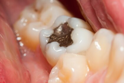
Dental X-rays are pictures of your teeth and the bones that support them. They help detect and diagnose a wide range of problems including cavities, bone loss due to gum disease, impacted or unerupted teeth, cysts, abscesses, and tumors. Before taking X-rays, we will place a lead apron over your chest and a thyroid collar around your neck to minimize your exposure to radiation.
Dental x-ray tampa fl Digital radiography uses electronic sensors to capture and display images of your teeth and surrounding structures on a monitor in the treatment room. These images can be enlarged and enhanced to help us better detect any problem areas. In addition, digital X-rays provide lower exposure levels (up to 90% less) than traditional film X-rays and require no chemical processing.
X-rays are an essential preventative diagnostic tool that reveal important information not visible during a visual examination. They allow us to safely and accurately detect hidden abnormalities such as tooth and jawbone structure, impacted or extra teeth, gum disease, bone loss, and other problems. Without X-rays, these problems may go undetected until they become more serious and costly to treat. X-rays are an integral part of your dental exam and necessary for a complete and accurate diagnosis. X-rays are safe, painless, and comfortable. There are no significant risks associated with X-rays when administered by a trained professional in the proper settings.
Intraoral X-rays Dental X-rays, sometimes called radiographs, are a valuable diagnostic tool used to identify and track problems with the teeth and jaws. They are an important component of comprehensive oral care and should be taken every 1 to 2 years. X-rays are safe and effective, and they only expose a small amount of radiation. X-rays can be taken inside (intraoral) or outside the mouth (extraoral). Intraoral X-rays show the entire tooth in one image and are usually taken at recall visits to detect new cavities, monitor tooth growth and development, and examine bone health. They are also used to evaluate the success of fillings and other treatments.
Extraoral X-rays are larger and capture the entire upper and lower jaw in one image. They are often needed for new patients and may be required before a dental treatment plan is developed. They can also be used to detect impacted or developing teeth, and to monitor jaw growth and development, as well as the status of sinuses, temporomandibular joints (TMJ), and tumors.
Bitewing X-rays Bitewing X-rays are the type of X-rays most patients are familiar with. They show both upper and lower teeth at one time, allowing your dentist to detect decay in between back teeth that is not visible with the naked eye. These X-rays also help diagnose gum disease and bone loss. With these X-rays, you will need to bite down on a small device that holds the sensor or film in place. You will need to remain as still as possible during the procedure to prevent blurred images. Your dentist will then activate the X-ray machine and position it at your mouth. Then, you will need to close your mouth and hold the sensor in place for a few seconds. To minimize radiation exposure, your dentist will cover you with a lead apron and thyroid collar. The X-ray will then capture an an image of your tooth and surrounding bone structure. This will help your dentist identify potential problems and treat them before they become serious.
Cone Beam Computerized Tomography (CBCT) Cone Beam Computerized Tomography (CBCT) is a revolutionary dental technology that enables us to obtain detailed 3D visuals of your mouth and jaw. It is a non-invasive imaging process that is used to enhance diagnostic accuracy, increase treatment planning efficiency, and ensure a comfortable patient experience. In a single rotation, the CBCT scanner will produce anywhere from 150 to 200 high-resolution two-dimensional (2-D) images that are then digitally combined to create a 3-D model of your skeletal and dental structures. It takes less than a minute to complete and reduces radiation exposure by 90% compared to traditional CT scans.
CBCT is critical for procedures like dental implants, orthodontics, and TMJ evaluations. It provides a precise assessment of bone structure, nerve pathways, and sinus cavities for effective treatment planning. It also helps to identify impacted or unruptured teeth, and assists in diagnosing cysts or tumors. In addition, it allows us to accurately evaluate the thickness and density of your jawbone to determine whether it is suitable for dental implant placement.
The advanced imaging capabilities of dental x-ray tampa fl also play a vital role in detecting subtle pathologies that might be missed with conventional X-rays, leading to earlier intervention and better outcomes. Furthermore, the detailed 3D models allow for virtual surgical planning, enabling dentists to precisely map out procedures and anticipate potential challenges, thereby increasing the predictability and success rate of complex treatments. This comprehensive insight into your oral anatomy ensures a highly personalized and effective approach to your dental health.